

Gram-Negative Bacteria
Gram-negative bacteria play a major role in biology, although many people don’t realise how frequently they influence human life and the environment. This structural difference clearly separates them from Gram-positive bacteria and also explains why many Gram-negative bacteria are harder to kill with antibiotics.
This article helps you understand what Gram-negative bacteria are, explains their cell wall composition, and highlights the features that make them different from other bacteria.
Table of Contents
- What Are Gram-Negative Bacteria
- Classification of Gram-Negative Bacteria
- Key Components of the Gram-Negative Cell Wall
- Gram-Negative Bacteria: When Do They Cause Infections
- Frequently Asked Questions on Gram-Negative Bacteria

What Are Gram-Negative Bacteria?
If you’ve ever noticed why some bacteria appear pink instead of purple during Gram staining, you’re actually looking at one of the biggest clues that identifies Gram-negative bacteria.

These microorganisms do not retain the crystal violet stain and instead turn pink/red with safranin, all because of their unique three-layered cell envelope.
You’ll find Gram-negative bacteria almost everywhere in soil, water, plants, the human body, and almost all environments that support life.
Some, like E. coli, are harmless or even helpful, while others, such as Pseudomonas, Klebsiella, or Neisseria, can cause serious infections.
Now, what makes them so distinct?
Their structure holds the answer.
-
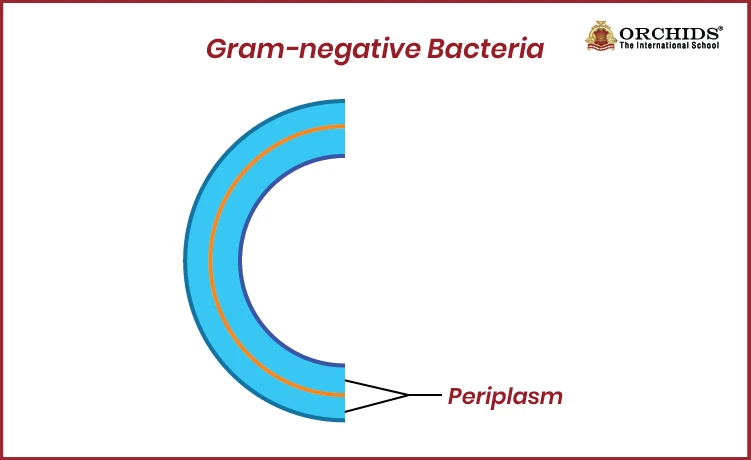
Gram-negative bacteria have a thin peptidoglycan layer sandwiched between an inner membrane and an outer membrane rich in lipopolysaccharides (LPS).
 \
\
-
They also contain porins for transport, a busy periplasmic space filled with enzymes, and Braun’s lipoprotein that anchors the outer membrane.
-
They lack teichoic acids, rarely form spores, and when flagella are present, they have four rings instead of two.
The cell envelope of gram-negative bacteria consists of three layers , but what does each layer actually do?
Let’s discuss.
1. The outer membrane is a specialised bilayer that acts as the first protective barrier, with phospholipids on the inner leaflet, LPS on the outer leaflet, porins for molecule entry, and Braun’s lipoprotein for stability.
2. Periplasmic space is a gel-like compartment between the membranes that holds enzymes for nutrient processing, transport proteins, and a thin peptidoglycan layer essential for metabolism and antibiotic resistance.
3. Cytoplasmic (Inner) membrane is a phospholipid bilayer that regulates nutrient transport, supports metabolic activities, and drives energy production within the cell.
All of these features work together to make Gram-negative bacteria more structurally complex and often more resistant to many antibiotics.
Also Read: Difference Between Gram-Positive and Gram-Negative Bacteria
Classification of Gram-Negative Bacteria
When you start understanding gram-negative bacteria, you’ll notice they can’t be grouped by just one feature.
Instead, scientists classify gram-positive and gram-negative bacteria based on several factors that help explain their structure, behaviour, and where they’re commonly found.
To begin with:
1. Gram-positive and gram-negative bacteria are identified by their Gram stain result; they appear pink or red after taking up the counterstain safranin. This is linked to their cell wall structure, which includes a thin peptidoglycan layer and an outer membrane.
2. Beyond structure, their habitat also plays an important role. Gram-negative bacteria may live in soil, water, or the human gut or act as pathogens that cause disease.
3. Some are opportunistic, infecting only when immunity is low, while others are strict pathogens that cause infections even in healthy individuals.
So, how does gram staining identifies gram-negative bacteria?
Let’s find out.
If you’re wondering how scientists distinguish these bacteria so quickly, the Gram staining process makes it surprisingly clear. The procedure uses four steps, such as crystal violet, iodine, alcohol or acetone, and finally safranin.
Gram-negative bacteria lose the crystal violet during the decolorisation step because their thin peptidoglycan layer cannot hold the dye.
Once this happens, safranin stains them pink, making them easy to tell apart from purple-stained gram-positive bacteria.
Key Components of the Gram-Negative Cell Wall
The gram-negative cell wall is more complex than that of gram-positive bacteria. Its unique multilayered structure, especially the presence of an outer membrane, gives these bacteria added protection, stronger antibiotic resistance, and distinct pathogenic features.
Here’s a clear breakdown of its key structural elements:
Gram-Negative Bacteria: When Do They Cause Infections?
It is interesting tp note that gram-negative bacteria are found everywhere around us in water, soil, food, hospital surfaces, and even inside the human body. Most of them stay harmless, but under the right conditions, they can quickly turn into major disease-causing pathogens.
1. Many infections begin with bacteria like E.coli, which normally live safely in the gut but can cause trouble when they enter places they don’t belong like the urinary tract leading to UTIs, gastroenteritis, food poisoning, or even neonatal meningitis.
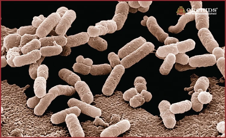
2. Others, such as Pseudomonas aeruginosa, thrive in moist environments and become problematic in hospitals.
They can infect wounds, lungs, or urinary tracts, especially in people with weak immunity.
3. They cause pneumonia, bloodstream infections, and severe wound infections, particularly in individuals on ventilators or catheters or recovering from injuries.
4. Neisseria gonorrhoeae is a prime example, spreading through unprotected sexual contact and leading to gonorrhoea.
5. Members of the Enterobacteriaceae family contaminate food and water, causing food poisoning, UTIs, and pneumonia.
A major concern today is the rise of carbapenem-resistant Enterobacteriaceae (CRE), superbugs that have developed extreme resistance to antibiotics, making infections difficult to treat and control.
In this article, we understood that together, these bacteria show how Gram-negative species, although everyday organisms, can turn opportunistic when the immune system weakens, when hygiene lapses occur, or when medical environments provide the perfect conditions for them to spread and cause serious disease.
Frequently Asked Questions on Gram-Negative Bacteria
1. Why is the LPS (Endotoxin) Layer Medically Important?
The LPS layer causes strong immune reactions like fever and inflammation and can lead to septic shock; when bacteria die, released endotoxins worsen the illness.
2. Why Are Gram-Negative Bacteria Often More Antibiotic-Resistant?
Their outer membrane blocks drugs, efflux pumps expel antibiotics, and enzymes deactivate medicines before they can work.
3. How Do Gram-Negative Bacteria Spread?
They spread through contaminated food and water, hospital equipment, unclean surfaces, direct contact, and inhalation of infected droplets.
4. Which Common Diseases Are Caused by Gram-Negative Bacteria?
Common Gram-negative bacteria examples include infections such as UTIs, pneumonia, food poisoning, bloodstream infections, and STDs like gonorrhoea.
5. Why Don’t Gram-Negative Bacteria Retain Crystal Violet Stain?
Because of their thin peptidoglycan layer and outer membrane, the dye cannot remain trapped. As a result, they appear pink when safranin is applied.
6. What Makes Gram-Negative Infections Hard to Treat?
Their layered cell structure limits drug entry, and frequent genetic mutations help them develop resistance faster than many other microbes.
7. Where Are Gram-Negative Bacteria Commonly Found?
They live in soil, water, the human gut, hospitals, and damp areas, making these Gram-negative bacteria examples hard to avoid and important to understand.
Science isn't just a subject, it's the way of seeing the world. Curious how Orchids The International School teaches it that way? Talk to our admissions team.
Admissions Open for 2026-27
What type of concept pages would you prefer?
CBSE Schools In Popular Cities
- CBSE Schools in Bangalore
- CBSE Schools in Mumbai
- CBSE Schools in Pune
- CBSE Schools in Hyderabad
- CBSE Schools in Chennai
- CBSE Schools in Gurgaon
- CBSE Schools in Kolkata
- CBSE Schools in Indore
- CBSE Schools in Sonipat
- CBSE Schools in Delhi
- CBSE Schools in Rohtak
- CBSE Schools in Bhopal
- CBSE Schools in Aurangabad
- CBSE Schools in Jabalpur
- CBSE Schools in Jaipur
- CBSE Schools in Jodhpur
- CBSE Schools in Nagpur
- CBSE Schools in Ahmednagar
- CBSE School In Tumkur











Orchids The International School is one of India's leading chains of CBSE and ICSE schools, with 110+ schools across the country.